
Abductor Pollicis Longus: Anatomy, Function, Clinical Insights | Althox
The human hand is a marvel of evolutionary engineering, capable of intricate movements and powerful grips, largely thanks to a complex network of muscles and tendons. Among these, the Abductor Pollicis Longus (APL) stands out as a pivotal player in thumb mobility. This muscle, often overlooked in general discussions, is fundamental for a wide range of daily activities, from writing and grasping objects to more complex tasks requiring fine motor control.
Understanding the APL's anatomy, function, and common pathologies is crucial for healthcare professionals, athletes, and anyone interested in the biomechanics of the upper limb. Its distinct role in thumb abduction and extension makes it a frequent subject of study in orthopedics and rehabilitation. This comprehensive guide delves into the specifics of this vital muscle, exploring its origins, insertions, actions, and clinical significance.
Anatomical Description of the Abductor Pollicis Longus (APL)
The Abductor Pollicis Longus (APL) is one of the extrinsic muscles of the hand, meaning its muscle belly is located in the forearm, but its tendons extend into the hand to act on the thumb. It belongs to the deep layer of the posterior compartment of the forearm, specifically within the first dorsal compartment of the wrist, alongside the Extensor Pollicis Brevis (EPB). This anatomical proximity is significant, as both tendons share a common synovial sheath, making them susceptible to similar inflammatory conditions.
The APL is a relatively long muscle, as its name "longus" suggests, and plays a crucial role in the precise movements of the thumb. Its strategic placement allows it to exert considerable leverage on the first metacarpal bone, facilitating powerful and controlled thumb abduction. This muscle is often palpable on the radial side of the wrist, especially when the thumb is actively abducted.
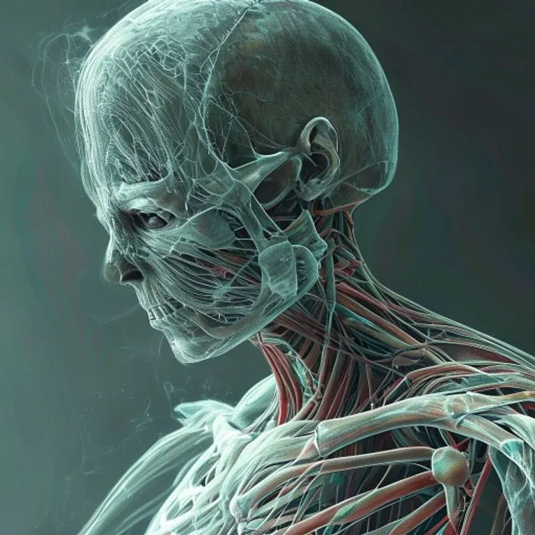
Anatomía detallada del Abductor Pollicis Longus, mostrando su origen, trayecto y la inserción en el pulgar.
Origin, Insertion, and Course
The Abductor Pollicis Longus muscle has a complex origin, arising from multiple points on the posterior surfaces of both the ulna and the radius, as well as the intervening interosseous membrane. Specifically, it originates from the lateral part of the dorsal surface of the ulna, distal to the insertion of the anconeus muscle. It also takes origin from the middle third of the dorsal surface of the radius and the adjacent interosseous membrane. This broad origin provides a stable base for its powerful actions on the thumb.
From its origin, the muscle fibers converge into a strong tendon that courses obliquely downwards and laterally across the forearm. This tendon then passes through the first dorsal compartment of the extensor retinaculum at the wrist, a fibrous band that holds tendons in place. Within this compartment, it is accompanied by the tendon of the Extensor Pollicis Brevis (EPB). This shared compartment is a critical anatomical detail, as it is often implicated in inflammatory conditions.
The primary insertion of the APL tendon is onto the radial side of the base of the first metacarpal bone. This direct attachment to the metacarpal allows the APL to effectively abduct and extend the thumb at the carpometacarpal (CMC) joint. However, anatomical variations are common, with accessory slips often inserting into the trapezium, the abductor pollicis brevis muscle, or even the thenar fascia. These variations can influence both the functional capabilities and the susceptibility to certain pathologies.
Functional Biomechanics
The primary action of the Abductor Pollicis Longus is the abduction of the thumb at the carpometacarpal joint. Thumb abduction is the movement of the thumb away from the palm, perpendicular to the plane of the palm. This motion is essential for grasping large objects and for the initial positioning of the thumb during various manipulative tasks. The APL also contributes to the extension of the thumb at the CMC joint, moving it posteriorly away from the other digits.
Beyond its direct actions on the thumb, the APL also acts as a weak abductor of the wrist, especially when the thumb is actively abducted. This synergistic action helps to stabilize the wrist during powerful gripping or pinching movements. The coordinated action of the APL with other thumb muscles, such as the Extensor Pollicis Brevis, Extensor Pollicis Longus, and the thenar muscles (Abductor Pollicis Brevis, Flexor Pollicis Brevis, Opponens Pollicis), allows for the wide range of complex thumb movements that are unique to humans.
The thumb's ability to abduct, flex, extend, and oppose the other fingers is critical for fine motor skills and dexterity. Without a properly functioning APL, tasks like holding a pen, typing, or even turning a doorknob can become significantly challenging. Its role in maintaining the thumb's position and facilitating its movement is indispensable for the overall functionality of the hand.
Innervation and Blood Supply
The Abductor Pollicis Longus muscle receives its nerve supply from the posterior interosseous nerve, which is a deep branch of the radial nerve. The radial nerve itself originates from the posterior cord of the brachial plexus (C5-T1 spinal nerves). The posterior interosseous nerve innervates most of the muscles in the posterior compartment of the forearm, controlling extension and supination movements. Damage to this nerve can significantly impair APL function, leading to weakness or paralysis of thumb abduction and extension.
The blood supply to the APL muscle is primarily provided by the posterior interosseous artery, which branches off the common interosseous artery, itself a branch of the ulnar artery. This arterial supply ensures adequate oxygen and nutrient delivery to the muscle, supporting its metabolic demands during activity. Understanding the neurovascular supply is vital for surgical procedures in the forearm and for diagnosing conditions that may affect muscle viability or function.
Clinical Significance and Pathologies
The Abductor Pollicis Longus is frequently involved in a common and often painful condition known as De Quervain's Tenosynovitis. This condition is characterized by inflammation and thickening of the synovial sheath that surrounds the tendons of the APL and EPB as they pass through the first dorsal compartment of the wrist. The narrowing of this compartment restricts the smooth gliding of the tendons, leading to pain, tenderness, and swelling on the radial side of the wrist, especially with thumb and wrist movements.
De Quervain's tenosynovitis is often associated with repetitive activities involving gripping, pinching, or wringing motions, such as those performed by new mothers (sometimes called "mommy thumb"), golfers, carpenters, or individuals who frequently use smartphones. The repetitive strain causes friction and irritation within the tendon sheath, perpetuating the inflammatory cycle. Early diagnosis and intervention are key to preventing chronic pain and functional limitations.
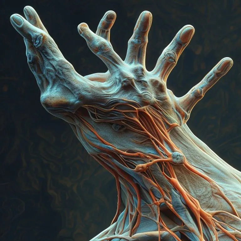
Herramientas clínicas para el diagnóstico y tratamiento de la tenosinovitis de De Quervain.
Diagnosis and Treatment of APL-Related Conditions
Diagnosing De Quervain's tenosynovitis typically involves a physical examination and a specific diagnostic test known as Finkelstein's test. During this test, the patient makes a fist with the thumb tucked inside the fingers, and then the wrist is ulnarly deviated (bent towards the little finger). A positive test elicits sharp pain along the radial side of the wrist, indicating inflammation of the APL and EPB tendons. Imaging studies, such as ultrasound or MRI, can confirm the diagnosis by visualizing tendon sheath thickening and inflammation, though they are often not necessary for typical cases.
Treatment for De Quervain's tenosynovitis usually begins with conservative measures. These include rest, activity modification to avoid aggravating movements, and the use of non-steroidal anti-inflammatory drugs (NSAIDs) to reduce pain and inflammation. Splinting the thumb and wrist in a neutral position can also provide significant relief by immobilizing the affected tendons and promoting healing. Corticosteroid injections into the tendon sheath are often highly effective in reducing inflammation and pain, providing longer-lasting relief than oral medications.
In cases where conservative treatments fail to provide adequate relief, surgical intervention may be considered. The surgical procedure, known as a De Quervain's release, involves incising the first dorsal compartment retinaculum to decompress the APL and EPB tendons. This widens the tunnel, allowing the tendons to glide freely without friction. Surgical outcomes are generally excellent, with most patients experiencing complete relief of symptoms and restoration of function.
Rehabilitation and Prevention Strategies
Following treatment for APL-related conditions, particularly De Quervain's tenosynovitis, rehabilitation plays a crucial role in restoring full hand and wrist function. Physical or occupational therapy often involves a progressive exercise program designed to improve range of motion, strength, and flexibility of the thumb and wrist. These exercises typically start with gentle passive and active movements, gradually advancing to strengthening exercises using resistance bands or light weights. Modalities such as ice, heat, and ultrasound may also be used to manage pain and inflammation.
Preventive measures are equally important, especially for individuals engaged in repetitive tasks. Ergonomic adjustments at work or home can significantly reduce strain on the APL and EPB tendons. This includes using proper posture, adjusting keyboard and mouse positions, and taking frequent breaks to stretch and rest the hands. Awareness of repetitive motions and modifying them to reduce stress on the wrist and thumb can help prevent the onset or recurrence of De Quervain's tenosynovitis. Education on proper body mechanics and activity modification is a cornerstone of prevention.
- Ergonomic Assessment: Evaluate workspaces and daily routines for repetitive strain risks.
- Activity Modification: Adjust techniques for tasks involving gripping, pinching, or wringing.
- Regular Stretching: Perform gentle stretches for the thumb, wrist, and forearm to maintain flexibility.
- Strengthening Exercises: Incorporate exercises to build strength in forearm and hand muscles.
- Rest Breaks: Take frequent short breaks during prolonged repetitive activities.
Anatomical Variations of the APL
Anatomical variations of the Abductor Pollicis Longus are quite common and can have clinical implications. The APL muscle and its tendon are known for their variability in terms of the number of muscle bellies, the number of tendons, and their insertion points. Studies have shown that the APL can have two or more tendons in a significant percentage of the population, often with accessory slips inserting into various structures beyond the first metacarpal, such as the trapezium, the thenar muscles (e.g., Abductor Pollicis Brevis), or the fascia overlying the thenar eminence.
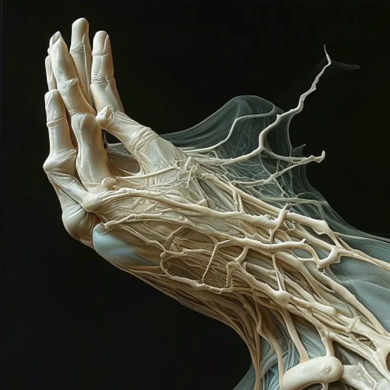
Representación conceptual del movimiento y la biomecánica muscular del pulgar.
These variations are particularly relevant in the context of De Quervain's tenosynovitis. The presence of multiple APL tendons or accessory slips within the already confined first dorsal compartment can predispose individuals to increased friction and inflammation, making them more susceptible to the condition. During surgical release, a surgeon must be aware of these potential variations to ensure complete decompression of all affected tendons and to prevent recurrence of symptoms. Pre-operative imaging, such as high-resolution ultrasound, can sometimes identify these anatomical peculiarities.
Understanding these anatomical nuances is crucial for both diagnostic accuracy and effective treatment. For instance, a patient with multiple APL tendons might experience more severe symptoms or require a more extensive surgical release than someone with a typical single tendon. Research continues to explore the prevalence and clinical impact of these variations, contributing to improved patient care and surgical techniques.
| Characteristic | Description |
|---|---|
| Location | Deep layer of posterior forearm compartment |
| Origin | Posterior surfaces of ulna, radius, and interosseous membrane |
| Insertion | Radial side of the base of the first metacarpal bone |
| Primary Action | Abduction of the thumb at the carpometacarpal joint |
| Secondary Action | Extension of the thumb at the CMC joint, weak wrist abduction |
| Innervation | Posterior interosseous nerve (branch of radial nerve, C5-T1) |
| Blood Supply | Posterior interosseous artery |
| Clinical Relevance | Commonly involved in De Quervain's tenosynovitis |
Conclusion
The Abductor Pollicis Longus muscle, though small in comparison to larger limb muscles, holds immense functional importance for the human hand. Its precise actions of thumb abduction and extension are indispensable for the dexterity and manipulative capabilities that define human interaction with the environment. From the simplest grasp to the most intricate fine motor tasks, the APL plays a silent yet critical role.
Furthermore, its susceptibility to conditions like De Quervain's tenosynovitis highlights its clinical relevance, affecting countless individuals who rely on their hands for work and daily life. A thorough understanding of its anatomy, biomechanics, and potential pathologies is essential for effective diagnosis, treatment, and rehabilitation. By appreciating the complexity and vital function of the APL, we gain deeper insight into the remarkable engineering of the human body and the importance of maintaining its health.
Fuente: Contenido híbrido asistido por IAs y supervisión editorial humana.


Comentarios