
Abductor Digiti Minimi: Anatomy, Function, Clinical Relevance | Althox
The abductor digiti minimi pedis, often referred to simply as the abductor of the little toe, is a critical intrinsic muscle of the foot. Despite its relatively small size, it plays a significant role in the biomechanics of the foot, particularly in maintaining balance and facilitating precise toe movements. Understanding its anatomy, function, and common pathologies is essential for both medical professionals and individuals seeking to comprehend the intricate workings of the human body.
This muscle is located along the lateral border of the foot, forming part of the plantar musculature. Its strategic position and attachments contribute to its primary actions: abduction and flexion of the fifth digit (the little toe). Beyond these direct actions, the abductor digiti minimi pedis is intimately involved in supporting the longitudinal arch of the foot and distributing pressure during gait, making it a silent workhorse in our daily mobility.
- Anatomical Overview: Origin, Insertion, and Innervation
- Functional Biomechanics: Role in Abduction, Flexion, and Arch Support
- Clinical Significance: Injuries and Pathologies
- Diagnosis and Treatment of Abductor Digiti Minimi Injuries
- Rehabilitation and Prevention Strategies
- Importance in Gait and Balance
- Comparative Anatomy: Abductor Digiti Minimi in Other Species
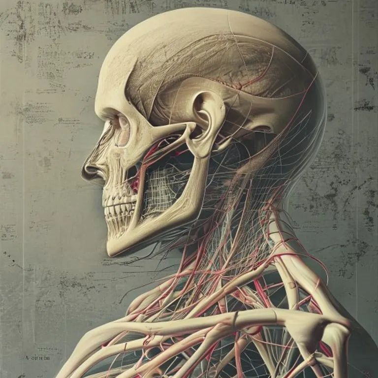
Detailed anatomical illustration of the abductor digiti minimi pedis, crucial for understanding its function.
Anatomical Overview: Origin, Insertion, and Innervation
The abductor digiti minimi pedis muscle is situated in the first layer of the plantar muscles, which are organized into four layers. Its robust structure is vital for the stability and movement of the lateral aspect of the foot. Understanding its precise anatomical attachments is fundamental to appreciating its biomechanical contributions.
The muscle primarily originates from several points on the calcaneus, the largest bone in the foot, commonly known as the heel bone. Specifically, it arises from the lateral process of the tuberosity of the calcaneus, a prominent bony projection. It also draws fibers from the plantar surface of the calcaneus, situated between the two processes of the tuberosity, and from the anterior aspect of the internal process of the plantar fascia. Furthermore, it receives contributions from the intermuscular septum that separates it from the flexor digitorum brevis, another muscle in the first plantar layer.
From its origin, the muscle fibers converge into a tendon that courses along the lateral side of the foot. This tendon then inserts into the fibular side (lateral side) of the base of the first phalanx of the fifth digit. It often shares its insertion point with the flexor digiti minimi brevis, highlighting a synergistic relationship between these muscles in controlling the little toe.
Innervation of the abductor digiti minimi pedis is provided by the lateral plantar nerve, which is a branch of the tibial nerve. The tibial nerve itself is a major nerve of the lower limb, originating from the sciatic nerve. This neural supply ensures that the muscle receives the necessary signals for contraction and proper function. Blood supply to this muscle is primarily from the lateral plantar artery, which branches from the posterior tibial artery, providing essential nutrients and oxygen for its metabolic demands.
Here is a summary of its key anatomical features:
- Origin: Lateral process of the tuberosity of the calcaneus, plantar surface of the calcaneus (between tuberosity processes), anterior part of the internal process of the plantar fascia, and intermuscular septum.
- Insertion: Fibular side of the base of the first phalanx of the fifth digit.
- Innervation: Lateral plantar nerve (branch of the tibial nerve).
- Blood Supply: Lateral plantar artery.
Functional Biomechanics: Role in Abduction, Flexion, and Arch Support
The primary functions of the abductor digiti minimi pedis are the abduction and flexion of the little toe. Abduction refers to the movement of the toe away from the midline of the foot, while flexion involves bending the toe downwards. Although these movements might seem minor, they are crucial for fine-tuning foot posture and adapting to uneven surfaces.
Beyond direct toe movement, this muscle plays a significant role in maintaining the structural integrity of the foot, particularly the lateral longitudinal arch. By connecting the calcaneus to the fifth metatarsal and phalanx, it acts as a dynamic stabilizer, helping to prevent the arch from collapsing during weight-bearing activities. This supportive role is often underestimated but is vital for efficient locomotion and shock absorption.
During the stance phase of gait, as the foot makes contact with the ground and bears weight, the abductor digiti minimi pedis contracts to stabilize the lateral side of the foot. This stabilization helps distribute plantar pressure evenly and provides a stable base for propulsion. In activities requiring quick changes in direction or balancing on one foot, the subtle actions of this muscle contribute significantly to overall stability and proprioception.
The synergistic action with the flexor digiti minimi brevis further enhances its effectiveness. While the abductor primarily abducts, the flexor primarily flexes. Together, they allow for a controlled and coordinated movement of the little toe, which, in turn, influences the broader mechanics of the forefoot. This intricate interplay underscores the complexity and efficiency of the foot's muscular system.
The intricate biomechanics of the foot, highlighting its role in balance and stability.
Clinical Significance: Injuries and Pathologies
Given its constant engagement in weight-bearing and movement, the abductor digiti minimi pedis is susceptible to various injuries and pathologies. These conditions can significantly impact foot function, leading to pain, instability, and altered gait patterns. Awareness of these issues is crucial for early diagnosis and effective management.
One common condition affecting this muscle is strain or overuse injury, particularly in individuals who engage in activities that place repetitive stress on the lateral foot. Athletes, dancers, and individuals who spend long hours standing or walking on hard surfaces are particularly vulnerable. Symptoms typically include localized pain along the lateral border of the foot, tenderness to touch, and pain exacerbated by toe movements or weight-bearing.
A specific pathology often associated with the abductor digiti minimi pedis is known as "weasel toe" or "orteil belette." This condition involves a painful inflammation or irritation of the muscle or its tendon, often resulting from chronic compression or friction. It is frequently observed in individuals who wear ill-fitting shoes, especially narrow footwear that compresses the little toe. Women are often more prone to this condition due to common footwear choices.
Furthermore, nerve entrapment of the lateral plantar nerve, which innervates this muscle, can lead to muscle weakness, atrophy, and sensory disturbances in the lateral foot. This can manifest as burning pain, numbness, or tingling sensations, and can severely impair the function of the abductor digiti minimi pedis, affecting both toe movement and foot stability.
In rare cases of polydactyly (the presence of an extra digit), the abductor digiti minimi pedis or a supernumerary muscle might be involved in the movement of the additional toe. This anatomical variation can sometimes lead to altered biomechanics and potential issues if not properly managed, although it is less common than overuse injuries.
Diagnosis and Treatment of Abductor Digiti Minimi Injuries
Diagnosing injuries to the abductor digiti minimi pedis typically involves a thorough clinical examination. A healthcare professional will assess the patient's symptoms, palpate the lateral aspect of the foot for tenderness, and evaluate the range of motion and strength of the little toe. Specific tests, such as resisted abduction and flexion of the fifth digit, can help pinpoint the affected muscle.
Imaging studies, such as ultrasound or Magnetic Resonance Imaging (MRI), may be used to confirm the diagnosis, particularly in cases of suspected tears, severe inflammation, or nerve entrapment. Ultrasound is often preferred for its ability to visualize soft tissues dynamically and its cost-effectiveness, while MRI provides more detailed anatomical information.
Treatment approaches vary depending on the severity and nature of the injury. Conservative management is usually the first line of treatment for strains and overuse injuries. This typically includes:
- Rest: Avoiding activities that aggravate the pain.
- Ice: Applying ice packs to reduce inflammation and pain.
- Compression: Using bandages or wraps to provide support and reduce swelling.
- Elevation: Raising the foot to minimize swelling.
- Anti-inflammatory medication: Over-the-counter non-steroidal anti-inflammatory drugs (NSAIDs) can help manage pain and inflammation.
- Footwear modification: Switching to wider, more supportive shoes to reduce compression on the little toe.
For persistent symptoms or more severe conditions like nerve entrapment, physical therapy plays a crucial role. Therapists may employ techniques such as manual therapy, stretching exercises to improve flexibility, and strengthening exercises to restore muscle function and stability. In rare and severe cases, surgical intervention might be considered, particularly for chronic nerve entrapment or extensive muscle tears that do not respond to conservative measures.
Understanding the subtle yet critical functions of foot muscles is key to overall well-being.
Rehabilitation and Prevention Strategies
Effective rehabilitation is vital for full recovery from abductor digiti minimi injuries and for preventing recurrence. A structured exercise program, often guided by a physical therapist, focuses on restoring strength, flexibility, and proprioception in the foot. Prevention strategies are equally important, especially for individuals at high risk.
Rehabilitation exercises may include:
- Toe Spreads: Actively spreading the little toe away from the other toes to strengthen the abductor muscle.
- Marble Pick-ups: Using the toes to pick up small objects like marbles, which helps improve intrinsic foot muscle coordination and strength.
- Towel Crunches: Placing a towel on the floor and using the toes to gather it towards the heel, targeting the plantar muscles.
- Resistance Band Exercises: Using a resistance band around the little toe to provide external resistance during abduction movements.
- Balance Exercises: Standing on one leg or using wobble boards to improve overall foot and ankle stability.
Prevention primarily revolves around proper footwear and maintaining good foot mechanics. Choosing shoes with a wide toe box that allows ample space for the toes to spread naturally can prevent compression and reduce the risk of "weasel toe." Avoiding high heels and excessively narrow shoes is also recommended. Regular stretching and strengthening exercises for the foot and ankle can enhance muscle resilience and reduce susceptibility to injury.
Athletes should pay particular attention to proper warm-up routines and gradual progression of training intensity. Incorporating foot-specific exercises into their regimen can significantly reduce the risk of overuse injuries. For individuals with structural foot abnormalities, custom orthotics may be beneficial in providing additional support and correcting biomechanical imbalances, thereby offloading stress from the abductor digiti minimi pedis.
Importance in Gait and Balance
The abductor digiti minimi pedis, along with other intrinsic foot muscles, plays a crucial role in the complex processes of gait and balance. Its contribution, though subtle, is indispensable for efficient locomotion and maintaining an upright posture. During walking, running, and standing, the foot acts as a dynamic platform that adapts to various terrains and distributes forces throughout the lower limb.
During the initial contact phase of gait, the foot pronates slightly to absorb shock. As the body moves over the foot, the foot then supinates, becoming a rigid lever for propulsion. The abductor digiti minimi pedis assists in stabilizing the lateral column of the foot during these transitions, ensuring that the foot maintains its structural integrity and efficiently transfers forces. Weakness or dysfunction of this muscle can lead to excessive pronation or instability, potentially contributing to conditions like plantar fasciitis or ankle sprains.
In terms of balance, the intrinsic foot muscles, including the abductor digiti minimi pedis, act as fine-tuners. They provide proprioceptive feedback to the brain, informing it about the foot's position and movement in space. This feedback is critical for making rapid adjustments to maintain equilibrium, especially on uneven surfaces or during dynamic activities. A strong and well-coordinated abductor digiti minimi pedis contributes to a more stable base of support, reducing the risk of falls, particularly in older adults.
Research indicates that strengthening intrinsic foot muscles can improve balance and reduce the incidence of lower limb injuries. Therefore, exercises targeting the abductor digiti minimi pedis are not just for rehabilitation but also for enhancing athletic performance and promoting overall foot health and stability throughout the lifespan.
Comparative Anatomy: Abductor Digiti Minimi in Other Species
While this article focuses on the human abductor digiti minimi pedis, it's insightful to briefly consider its presence and function in other species. Comparative anatomy often reveals evolutionary adaptations and shared physiological principles across the animal kingdom. The presence and development of this muscle vary significantly depending on the species' mode of locomotion and foot structure.
In many quadrupedal mammals, the foot (or paw) serves different functions than in humans, often being adapted for running, climbing, or grasping. The intrinsic foot muscles in these animals are typically well-developed to provide strong grip, shock absorption, and propulsion. For instance, in primates, the abductor digiti minimi (both in hand and foot) can be highly developed, contributing to grasping capabilities, which are essential for arboreal locomotion.
In species with highly specialized feet, such as ungulates (hoofed animals), the individual toe muscles may be reduced or fused, as their locomotion relies on a single, strong digit. Conversely, in animals that use their feet for manipulation or fine motor control, like some rodents or marsupials, the intrinsic muscles, including abductors, might be more distinct and versatile.
Studying these variations helps us understand the evolutionary pressures that have shaped human foot anatomy and the specific adaptations that allow for bipedalism. The human abductor digiti minimi pedis, while seemingly small, is a testament to the intricate design of the human foot, optimized for upright posture, balance, and efficient terrestrial locomotion.
Fuente: Contenido híbrido asistido por IAs y supervisión editorial humana.
Comentarios