
Abdomen Anatomy: Comprehensive Guide to Core Health | Althox
The abdomen, a critical region in both human and animal anatomy, serves as a central hub for numerous vital physiological processes. Located between the chest and the pelvis, it houses a complex array of organs responsible for digestion, excretion, and reproduction, while also providing structural support and facilitating movement. Understanding the intricate architecture and diverse functions of the abdomen is fundamental to comprehending overall biological health and performance.
This comprehensive guide delves into the multifaceted aspects of the abdomen, exploring its anatomical definitions across different species, detailing the specific organs it contains, and examining the crucial role of its muscular wall. We will also discuss the importance of abdominal health, including the benefits of targeted exercise and the significance of surface landmarks in clinical assessment. From the macroscopic view of its boundaries to the microscopic functions of its cellular components, the abdomen is a marvel of biological engineering.
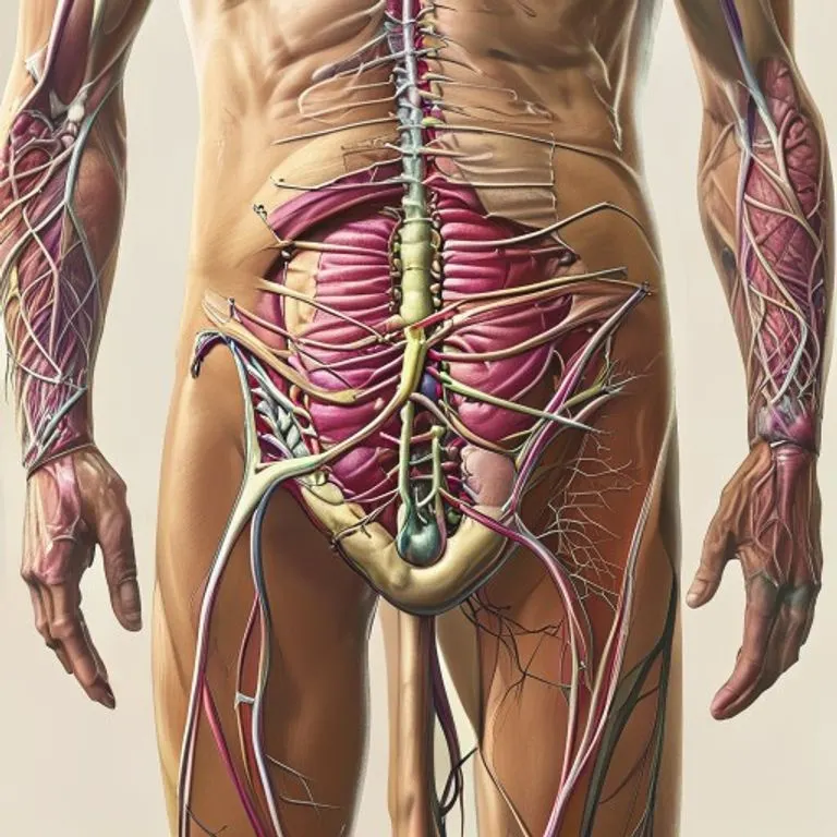
The human abdomen, a complex region, houses vital organs and muscular structures essential for life.
Table of Contents
- Defining the Abdomen: A Biological Overview
- Human Abdomen: Anatomy and Organ Systems
- The Muscular Architecture of the Abdomen
- Abdominal Health and Exercise
- The Invertebrate Abdomen: Diversity in Structure
- Surface Landmarks and Clinical Significance
- Conclusion: The Abdomen's Central Role
Defining the Abdomen: A Biological Overview
The term "abdomen" refers to the body region situated between the thorax (chest) and the pelvis. This definition holds true for a vast array of animals, encompassing both vertebrates and invertebrates, though its specific structural characteristics and contents vary significantly across species. In essence, it is a flexible and protective compartment designed to house and support crucial internal organs.
In vertebrates, such as mammals, the abdomen, often referred to as the "belly," forms the lower part of the trunk. It is separated from the thoracic cavity by the muscular diaphragm, a key structure in respiration. Below, it is continuous with the pelvic cavity, forming a larger abdominopelvic cavity in many anatomical descriptions. This region is critical for digestion, nutrient absorption, waste elimination, and in many cases, reproduction.
For invertebrates, particularly arthropods like insects, the abdomen represents the posterior segment of the body, typically located behind the thorax or cephalothorax. While it serves similar functional roles in housing digestive and reproductive systems, its external structure, often composed of segmented plates, differs markedly from its vertebrate counterpart. This diversity underscores the evolutionary adaptations of life forms to their environments and physiological needs.
Human Abdomen: Anatomy and Organ Systems
The human abdomen is a meticulously organized region, bounded superiorly by the thoracic diaphragm and inferiorly by the pelvic inlet, which extends from the lumbosacral angle (the intervertebral disk between L5 and S1) to the pubic symphysis and the superior edge of the pelvis. This area encloses the abdominal cavity, a large space lined by a serous membrane known as the parietal peritoneum, which is continuous with the visceral peritoneum covering the organs themselves.
Within this cavity, a multitude of organs work in concert to sustain life. The digestive system constitutes a significant portion of the abdominal contents, including the stomach, small intestine (duodenum, jejunum, ileum), and large intestine (cecum, appendix, ascending, transverse, descending, and sigmoid colon, and rectum). These organs are responsible for the breakdown of food, absorption of nutrients, and formation of waste.
Accessory digestive organs, such as the liver, gallbladder, and pancreas, are also vital residents of the abdomen. The liver performs detoxification, protein synthesis, and bile production; the gallbladder stores bile; and the pancreas produces digestive enzymes and hormones like insulin. The urinary system is represented by the kidneys and ureters, which are retroperitoneal (located behind the peritoneum) but functionally associated with the abdominal region, filtering blood and producing urine.
- Digestive Organs: Stomach, small intestine, large intestine, cecum, appendix.
- Accessory Digestive Organs: Liver, gallbladder, pancreas.
- Urinary System Components: Kidneys, ureters (retroperitoneal).
- Other Vital Organs: Spleen (involved in immune function and blood filtration), adrenal glands.
The abdominal wall, a complex structure of muscles, fascia, and skin, provides protection and support to these delicate organs. It is broadly divided into anterior (front), lateral (sides), and posterior (back) regions. The integrity of this wall is crucial for maintaining intra-abdominal pressure, assisting in respiration, and enabling trunk movement. The peritoneum, a thin, transparent membrane, not only lines the cavity but also forms folds (mesenteries) that suspend and support many of the abdominal organs, facilitating their blood supply and innervation.
The Muscular Architecture of the Abdomen
The muscles of the abdomen are a sophisticated group that provides structural integrity, facilitates movement, and protects the internal viscera. These muscles are often collectively referred to as the "core" and are essential for almost every physical activity, from standing upright to performing complex athletic maneuvers. They work synergistically to provide stability and mobility to the trunk.
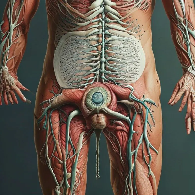
Detailed anatomical charts are vital tools for understanding the complex structures of the abdomen.
The primary abdominal muscles include the rectus abdominis, external obliques, internal obliques, and transversus abdominis. Each plays a distinct yet interconnected role in the overall function of the core. The rectus abdominis, often visible as the "six-pack" in well-conditioned individuals, runs vertically along the front of the abdomen, responsible for flexing the vertebral column (bending forward).
The external and internal obliques are located on the sides of the abdomen. The external obliques are superficial, with fibers running inferomedially, while the internal obliques lie beneath them, with fibers running superomedially. These muscles are crucial for trunk rotation, lateral flexion (bending sideways), and maintaining posture. Their coordinated action allows for dynamic and controlled movements of the torso.
The transversus abdominis is the deepest of the abdominal muscles, wrapping horizontally around the trunk like a corset. Its primary function is to compress the abdominal contents, increasing intra-abdominal pressure, which is essential for forced expiration, coughing, sneezing, and stabilizing the lumbar spine. This muscle is often considered the most important for core stability and postural support, despite not being externally visible.
| Muscle Group | Location | Primary Functions |
|---|---|---|
| Rectus Abdominis | Anterior surface, from pubis to ribs 5-7 | Flexes vertebral column, compresses abdominal viscera. |
| External Obliques | Lateral and anterior surfaces, superficial | Trunk rotation, lateral flexion, compresses abdomen. |
| Internal Obliques | Lateral and anterior surfaces, deep to external obliques | Trunk rotation, lateral flexion, compresses abdomen. |
| Transversus Abdominis | Deepest layer, encircles abdomen horizontally | Compresses abdominal contents, stabilizes lumbar spine. |
| Pyramidalis | Small, triangular muscle in lower abdomen (not always present) | Tenses linea alba. |
The coordinated action of these muscles is vital for maintaining proper posture, supporting the spine, and enabling efficient movement. Weak or imbalanced abdominal muscles can lead to various issues, including lower back pain, poor posture, and increased risk of injury. Therefore, strengthening these muscles is a cornerstone of physical therapy and fitness regimens.
Abdominal Health and Exercise
Maintaining a strong and healthy abdomen is paramount for overall physical well-being. Beyond aesthetic considerations, robust abdominal muscles contribute significantly to spinal stability, reduce the incidence of back pain, improve balance, and enhance athletic performance. A strong core acts as a natural corset, protecting internal organs and supporting the entire trunk.
Exercise plays a critical role in developing and maintaining abdominal strength. Various disciplines and specific exercises target different aspects of the abdominal musculature. Pilates and yoga, for instance, emphasize core engagement and stability through controlled movements and breathwork, improving both strength and flexibility. These practices often focus on engaging the deeper transversus abdominis and internal obliques, which are crucial for foundational core strength.
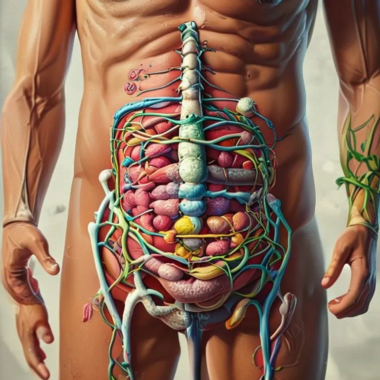
Conceptual art illustrating the intricate and dynamic metabolic processes within the abdominal region.
Traditional abdominal exercises, such as crunches and sit-ups, primarily target the rectus abdominis. Variations like bicycle crunches engage both the rectus abdominis and the obliques, contributing to a more comprehensive workout. However, it's crucial to perform these exercises with proper form to avoid strain on the neck or lower back. Incorporating exercises that challenge stability, such as planks and side planks, is also vital for strengthening the entire core unit.
Beyond bodyweight exercises, various fitness equipment can enhance abdominal training. The captain's chair, commonly found in gyms, allows for leg raises that effectively target the lower rectus abdominis and hip flexors. Stability balls are excellent tools for engaging core stabilizers, as they introduce an element of instability, forcing deeper muscles to activate. Suspension training systems, like TRX, offer advanced options for challenging core strength and endurance by leveraging body weight.
- Improved Posture: Strong abdominal muscles help maintain proper spinal alignment.
- Reduced Back Pain: A robust core supports the lumbar spine, alleviating pressure and discomfort.
- Enhanced Balance and Stability: Essential for everyday activities and athletic performance.
- Injury Prevention: A stable core reduces the risk of strains and sprains during physical exertion.
- Organ Protection: The muscular wall provides a protective barrier for internal organs.
It is important to remember that abdominal exercise should be part of a balanced fitness routine that also includes back strengthening, flexibility, and cardiovascular training. Over-focusing solely on "abs" without addressing other muscle groups can lead to imbalances and potential issues. A holistic approach to fitness ensures optimal abdominal health and overall physical resilience.
The Invertebrate Abdomen: Diversity in Structure
While the human abdomen is characterized by its muscular wall and internal cavity, the abdomen in invertebrates, particularly arthropods, exhibits a fascinating diversity of structures and adaptations. Despite these differences, its fundamental role in housing digestive and reproductive systems remains consistent across the animal kingdom. The invertebrate abdomen is typically segmented, reflecting the metameric body plan common to many arthropods.
In insects, the abdomen is generally composed of a series of segments, often numbering eleven, though this can vary significantly. Each segment is typically protected by hardened plates: a dorsal plate called a tergite and a ventral plate called a sternite. These plates are connected by flexible membranes, allowing for movement and expansion, which is crucial for processes like respiration, digestion, and egg-laying.
The internal contents of the insect abdomen include the digestive tract, which processes food and absorbs nutrients, and the reproductive organs, which are essential for species propagation. Unlike other arthropods, adult insects generally lack legs on their abdominal segments, though some primitive insects, like Archaeognatha, may possess rudimentary appendages. Many insect larvae, such as caterpillars, have fleshy prolegs on their abdominal segments, which aid in locomotion and gripping surfaces.
A notable example of abdominal modification can be seen in ants. In many ant species, the first segment of the abdomen is fused with the thorax to form a structure called the propodeum. The second segment often forms a narrow constriction known as the petiole, which can be followed by an additional segment, the postpetiole. The remaining segments form the bulbous gaster, which contains most of the internal organs. This specialized abdominal structure allows for greater flexibility and mobility, crucial for their complex social behaviors.
Other invertebrates, such as crustaceans and arachnids, also possess distinct abdominal regions with unique adaptations. For example, the abdomen of many crustaceans is muscular and used for swimming, while in spiders, it houses silk glands and respiratory organs. These variations highlight the remarkable evolutionary plasticity of the abdominal region in response to diverse ecological niches and lifestyles.
Surface Landmarks and Clinical Significance
The external surface of the human abdomen provides a wealth of anatomical landmarks that are invaluable in clinical assessment, diagnosis, and surgical planning. These visible or palpable features allow healthcare professionals to accurately locate underlying organs, identify areas of pain or swelling, and monitor physiological changes. Understanding these landmarks is fundamental for both medical practitioners and those seeking to understand their own body's topography.
In the midline of the anterior abdomen, a slight furrow, known as the linea alba, extends from the xiphoid process (the inferior tip of the sternum) down to the pubic symphysis. This fibrous band represents the fusion of the aponeuroses of the abdominal muscles. Approximately halfway along the linea alba lies the umbilicus, or navel, a prominent and easily identifiable landmark. The rectus abdominis muscles, when well-developed, can be seen on either side of the linea alba, often segmented by three or more transverse tendinous intersections, which contribute to the "six-pack" appearance.
The superior boundary of the abdomen is marked by the subcostal margin, formed by the cartilages of the 8th, 9th, and 10th ribs, which join to form a continuous arch. Inferiorly, the lateral abdominal muscles extend to the iliac crests of the hip bones and the inguinal ligaments, which run from the anterior superior iliac spine to the pubic tubercle. These lower boundaries are often marked by visible grooves in lean individuals, particularly those with high muscle definition.
Just above the pubic spines, on either side, are the superficial inguinal rings, which are openings in the muscular wall of the abdomen. In males, the spermatic cord passes through these rings; in both sexes, these are potential sites for inguinal hernias, where abdominal contents protrude through a weakened area of the wall. Palpation of these areas is a common part of physical examinations.
For more precise localization of internal structures, clinicians often divide the abdomen into regions or quadrants using imaginary lines. The most common method involves drawing two horizontal and two vertical lines. The horizontal lines include the transpyloric plane (Addison's line), located midway between the suprasternal notch and the top of the pubic symphysis, and the subcostal plane, connecting the lowest points of the tenth costal cartilages. The vertical lines are typically the midclavicular lines, extending downwards from the midpoints of the clavicles.
These divisions create nine distinct regions: epigastric, umbilical, hypogastric (pubic), right and left hypochondriac, right and left lumbar (flank), and right and left iliac (inguinal). Alternatively, a simpler system divides the abdomen into four quadrants: right upper, left upper, right lower, and left lower, using a horizontal line through the umbilicus and a vertical line along the linea alba. These regional classifications are crucial for documenting pain, identifying organomegaly (enlarged organs), and guiding diagnostic procedures.
Conclusion: The Abdomen's Central Role
The abdomen, whether in humans or other animals, stands as a testament to the complexity and efficiency of biological design. It is far more than just a physical space; it is a dynamic region where life-sustaining processes unfold, from the intricate dance of digestion and nutrient absorption to the critical functions of waste elimination and reproduction. Its robust muscular wall provides essential protection and stability, enabling movement and supporting the entire trunk.
From a human perspective, the health of the abdomen is inextricably linked to overall well-being. A strong core not only contributes to physical strength and athletic prowess but also plays a pivotal role in preventing common ailments like back pain and improving postural alignment. The diverse anatomical landmarks on its surface serve as vital guides for medical professionals, facilitating accurate diagnosis and treatment. Invertebrates, too, showcase the abdomen's adaptability, with segmented structures and specialized appendages tailored to their unique biological needs.
In conclusion, the abdomen is a central and indispensable component of the body plan, embodying a remarkable integration of form and function. Its study continues to provide profound insights into anatomy, physiology, and evolutionary biology, underscoring its enduring significance in the biological sciences and its critical importance for health across the animal kingdom. A holistic understanding and care for this vital region are fundamental for a healthy and active life.
Source: Hybrid content assisted by AIs and human editorial supervision.


Comentarios